Research
Macrophage metabolism in the pleural cavity
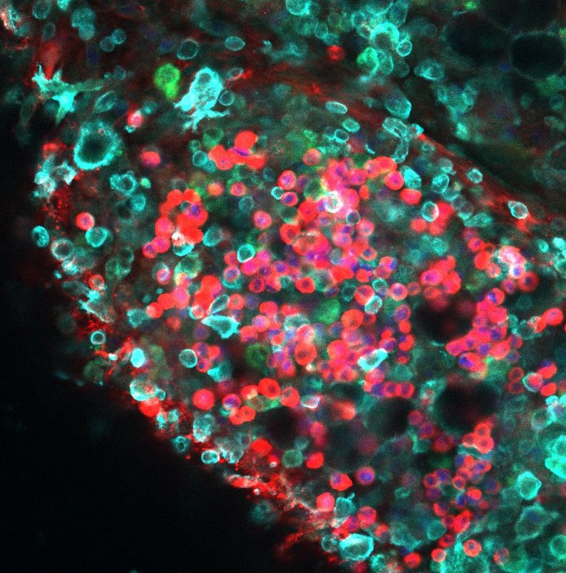
Internal organs are contained within three fluid-filled serous cavities: the peritoneum, which envelops the abdominal viscera; the pleural cavity enclosing the lungs and the pericardium which protects and anchors the heart. Fluid is secreted by the mesothelium lining the serosal membranes ensuring lubrication of the serous cavities. Serous fluid contains resident macrophages known as large cavity macrophages (LCM).
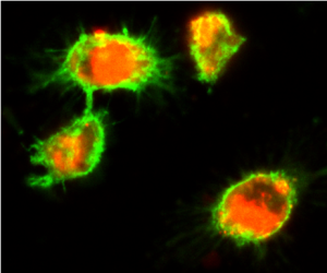
Following exposure to IL-4, either delivered exogenously or induced due to infection or type-2 inflammation, LCM will undergo a programme of M(IL-4) induced activation and proliferation increasing their number rapidly over the duration of the IL-4 exposure. Dedicated immunological niches called fat-associated lymphoid clusters (FALCs) develop under the intercellular pores formed by stomata in the mesothelium of specialised adipose tissues within the serous cavities. FALCs are sites of local IgM production during infection & inflammation. Our lab is interested in whether IgM is a co-factor for pleural LCM activation & division within the pleural cavity during Type-2 mediated inflammation. We are also investigating how LCM from the serous cavities utilise cholesterol, and what role oxidised phospholipids play in LCM biology during inflammation.
This work was funded by the Wellcome Trust and the MRC.
~~~Please see additional pages below for our other research projects ~~~