Laser Wakefield Accelerators (LWFA)
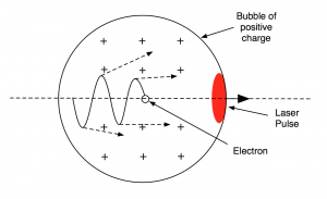
Laser driven plasma wakefields can be used to accelerate bunches of electrons to high energies over very small distances. A short, intense laser pulse incident on a gas target displaces electrons within the target creating regions of positive charge. This charge imbalance can be described as a plasma wave propagating in the wake of the laser. If the amplitude of this plasma wave, or wakefield, is great enough, electrons trapped in certain regions will be accelerated. This setup is much more compact than traditional particle accelerators, with the possibility to house such technology in university laboratories. Due to the large amount of energy electrons can gain in such a small distance, this type of accelerator can be utilised industrially to create high energy radiation such as x rays, which are used for high resolution imaging.
Accelerating Radiation
When electrons are accelerated, they emit electromagnetic radiation which is doppler shifted to high frequencies in the direction tangential to their motion. Using this trait, electrons oscillating periodically can be used as a light source. Most of the radiation is given off at the turning points of the orbit as that is when the acceleration is at its maximum. The figure below shows this mechanism [1].
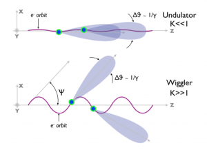
Radiation source which exploit this are called undulators or wigglers, depending on something known as the K parameter, which is a function of the period and the magnitude of the oscillation, as well as the particle energy. An undulator has K ≤ 1 and wiggler radiation has K > 1. Undulator radiation is emitted along the laser axis with a very small angular spread in one fundamental harmonic, whereas the direction of the wiggler radiation varies depending on the position of the electron in its orbit. A distant observer on the axis would see wiggler sourced light flashing on and off for this reason. Wiggler radiation can have a broad spectrum of harmonics with different frequencies. The characteristics of this spectrum are dependent on the K parameter.
Plasma Wiggler
As well as being accelerated in the direction of the laser, electrons oscillate transversely to the propagation of the laser due to electric and magnetic fields formed in the plasma [2]. These are known as betatron oscillations and are depicted in the figure below, with the dotted arrows illustrating emitted radiation.
Current experiments emit wiggler radiation. X-rays created in this way are essentially a by product of the acceleration process but have a number of useful properties which can be exploited for high resolution imaging purposes. The oscillation frequency, which determines the spectra of radiation given off via this mechanism, is higher than the frequency of x-rays generated by most conventional methods. This relates to a greater number of high energy photons per beam being produced. The duration of the radiation given off can be on the scale of a few femtoseconds since it is dependent on the size of the accelerated electron bunch. This is useful for viewing dynamic processes which take place on a small time scale. Also, it can be synchronised to a high precision with the laser in order to perform pump-probe measurements. This is where the laser (pump) collides with a material in order to excite or ionise it, and the x-ray (probe) beam follows shortly after to measure the new state.
Conventional Undulators/Wigglers
The emission process can be extended by a conventional undulator. This is a configuration of magnets, generally on the order of a meter in length, which create a periodic magnetic field.
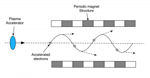
The high energy electron beam from the LWFA can be directed towards this structure, where the magnetic field will promote the transverse oscillations which lead to emission of synchrotron radiation. This technique can be applied to create extremely bright beams through FEL amplification. This is where an electron beam is bunched so close together that the radiation emitted by each electron constructively interferes with radiation emitted by other electrons in the bunch.
Electromagnetic Undulators/Wigglers
Another way to incite these transverse oscillations is using an electromagnetic undulator. This takes advantage of a process called Thomson Backscattering in order to produce high energy radiation. A second laser is fired at the laser plasma accelerated electron bunch in the opposite direction to their motion.
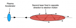
Interaction with the counter propagating electromagnetic wave causes the electrons to oscillate and in turn emit high frequency radiation in the forward direction. Although this is essentially very similar to the previous two undulator processes, the emitted radiation can be of higher energy, with the possibility of gamma ray emission.
Decelerating Radiation Sources
When high energy electrons interact with nuclei they lose energy and give off Bremsstrahlung or “braking radiation”. If an electron beam of high enough energy is directed at a target capable of decelerating the electrons quickly, this radiation can be in the x-ray, or even gamma spectrum. This mechanism is exploited by common commercial devices such as micro focus x-ray tubes, with the electron beam being formed by accelerating electrons across a potential difference. Micro-CT scanners use x-rays formed in this way to take reasonably high resolution pictures, which can be reconstructed to create 3D images of bones. Although resolution is limited by the energy of the accelerated electrons, these scanners are relatively affordable, practical imaging devices which can easily be implemented industrially and for medical purposes.
For future sources, the laser plasma acceleration method discussed earlier could be used to create a high quality electron beam to fire at the target. This could produce radiation with little divergence from the axis, capable of creating higher resolution images than current micro-CT scanners. Also, the bunch duration can be very small, meaning the radiation created can image processes which take place over small time periods.
Comparison of Techniques
At present, x ray radiation for imaging and probing materials tend to be provided by synchrotron accelerators or micro focus x-ray tubes. Synchrotrons either use bending magnets or conventional magnetic undulators to incite electrons to produce this radiation. To demonstrate the scale of a high energy synchrotron experiment, the picture below illustrates the size of the Diamond Light Source in Oxfordshire [3]. The building is roughly the size of six football pitches.

The energy of the light emitted and its average brilliance can be very high using an accelerator of this size, meaning it is capable of performing high resolution imaging. Micro focus tubes are on the opposite end of the spectrum as they are much more accessible, but have a comparatively low image resolution (although still pretty good for most purposes). Laser plasma x-ray sources hope to initially fill the void between these two mainstream sources, before potentially competing with synchrotron sources on resolution when the technology is better understood.
The table below shows a comparison between the theoretical characteristics of accelerating radiation from laser plasma sources with an electron energy of 1 GeV and the Diamond light source radiation properties from 3 GeV electrons. LWFA x ray sources compare favourably in terms of spatial and temporal resolution (source size and pulse duration), peak brilliance and energy as well as having the ability to synchronise to a pump laser with high accuracy. The low rep rate of the laser though is the factor which limits the average brilliance.
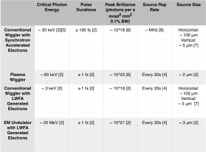
The sources that use the Bremsstrahlung process are difficult to compare using the same parameters. They produce a spectrum of photons which have a maximum energy equal to the electrons beam energy but the average emission energy is significantly lower than that. The characteristics of the spectrum are determined by the target material and shape. Micro focus tubes are a continuous source which usually aim to have peak photon energies between 15keV and 50keV [4]. Laser plasma accelerated electrons incident on a target can create the signature short pulse duration, with potential photon energies well into the MeV range. These high energy gamma photons are less useful for imaging purposes but can be used for cancer treatment if an intense beam can be formed.
All of these laser plasma x ray sources will benefit from the constant improvement of laser technology and from researchers forming a better experimental understanding of the wakefield acceleration regime. In the near future though, these techniques could easily be implemented for medical and industrial purposes on a wider scale.
References
[1] Kneip, S, (2010). Laser plasma accelerator and wiggler, Imperial College
[2] Corde, S., et al, (2013). Femtosecond x rays from laser-plasma accelerators. Reviews of Modern Physics, 85(1), pp.1-48.
[3] http://www.diamond.ac.uk/Home/About
[4] Cole, J., et al, (2015). Laser-wakefield accelerators as hard x-ray sources for 3D medical imaging of human bone. Scientific Reports, 5(1).
[5] Materlik G, Rayment T, Stuart DI. 2015. Diamond Light Source: status and perspectives (Introduction). Phil. Trans. R. Soc. A 373, 20130161
[6]Kneip, S., Najmudin, Z. and Thomas, A. (2012). A plasma wiggler beamline for 100 TW to 10 PW lasers. High Energy Density Physics, 8(2), pp.133-140.
[7] http://www.diamond.ac.uk/Science/Machine.html
[8] Chubar, O., et al. (2016). Ultra-high-resolution inelastic X-ray scattering at high-repetition-rate self-seeded X-ray free-electron lasers. Journal of Synchrotron Radiation, 23(2), pp.410-424.