We develop and apply a family of genetically encoded cell cycle biosensors based on the Fucci platform developed by Prof. Atsushi Miyawaki and Dr Asako Sakaue-Sawano (RIKEN). Our biosensors enable real-time visualisation of cell cycle dynamics in live cells, tissues and whole organisms.
Our biosensor family
We have developed five biosensor variants, each optimised for different experimental contexts:
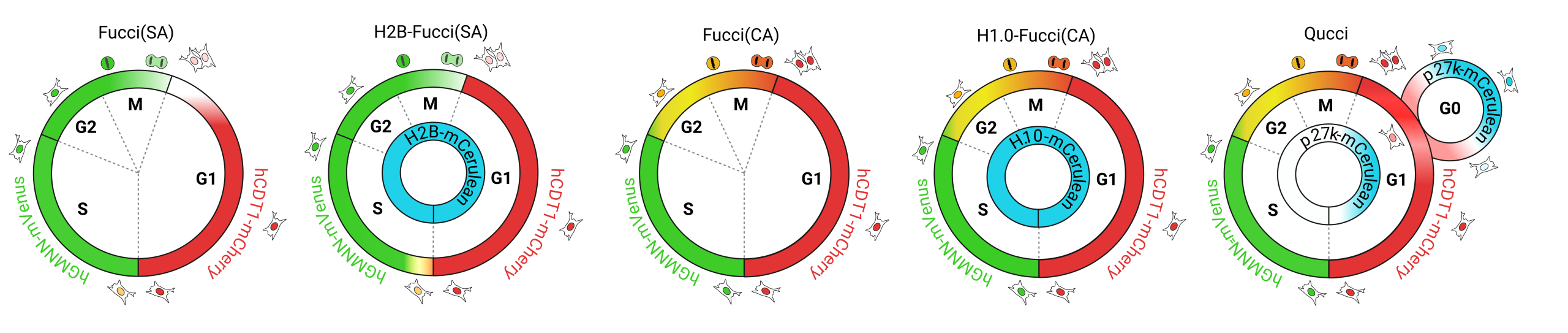
- Fucci(SA) — the original Fucci2a bicistronic biosensor, marking G1 in red and S/G2/M in green (Mort et al. Cell Cycle 2014)
- H2B-Fucci(SA) — tricistronic version incorporating H2B-Cerulean for precise single-cell tracking (Loftus et al. Dev Cell 2025)
- Fucci(CA) — the latest Fucci(CA) variant as a bicistronic construct marking G1, S, G2/M (Sakaue-Sawano et al. Mol Cell 2017)
- H1.0-Fucci(CA) — tricistronic version of Fucci(CA) incorporating H1.0-Cerulean for improved single cell tracking (Sudderick et al. bioRxiv 2026)
- Qucci — our new extended tricistronic cell cycle biosensor incorporating a p27k−Cerulean reporter to mark quiescent G0 cells alongside cycling populations
How they work
Fucci biosensors exploit the oscillating abundance of cell cycle-regulated proteins to mark distinct phases with spectrally separable fluorescent reporters.
Fucci(SA)
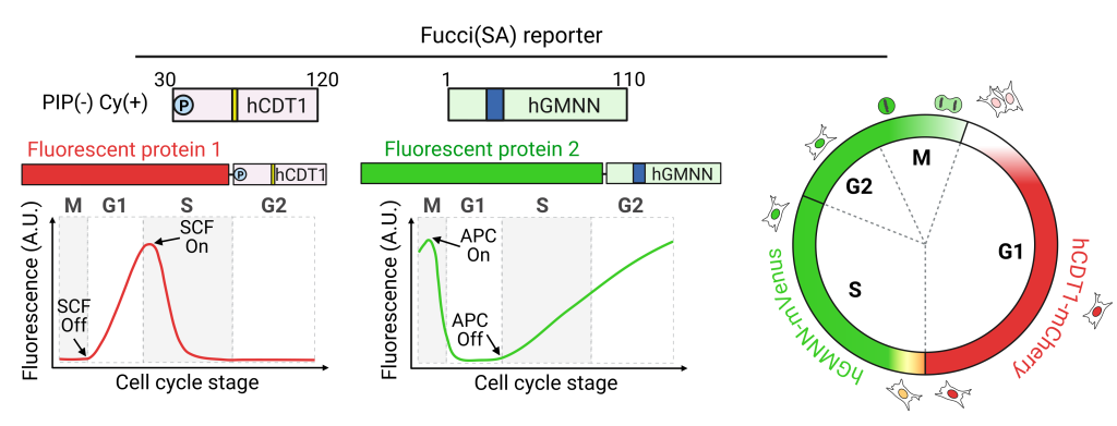
The original Fucci(SA) probe pair comprises a fusion of monomeric Kusabira Orange (mKO2 – red) with a truncated human CDT1 (amino acids 30–120), and a fusion of monomeric Azami Green (mAG – green) with the N-terminal 110 amino acids of human Geminin (GMNN). The red probe accumulates in G1 phase, labelling nuclei red, and is degraded at the G1–S transition via SCFSkp2. The green probe accumulates during S, G2 and M phases, labelling nuclei green, and is rapidly degraded before cytokinesis via APC/C.
An improved version of Fucci(SA) – replaces mKO2 and mAG with mCherry and mVenus respectively, increasing signal separation and compatibility with other reporters. Incorporation of a 2A self-cleaving peptide enabled equimolar co-expression of both reporters from a single promoter (Fucci2a or tandem-Fucci(SA)), facilitating the development of Cre-inducible mouse models.
Fucci(CA)
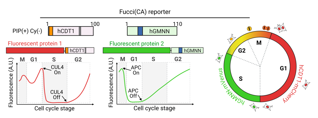
The tandem-Fucci(CA) probe pair updates the CDT1 probe from Fucci(SA) by removing the SCFSkp2-targeted Cy motif and instead incorporating an N-terminal PIP box, which targets CDT1 for degradation in S-phase via CUL4Ddb1. This results in CDT1 accumulation from the S–G2 transition through G2, M and G1, with rapid degradation at the G1/S boundary. With just two sensors, Fucci(CA) labels G1-phase nuclei red, S-phase nuclei green, and G2/M nuclei yellow.
Mouse lines
The R26Fucci2aR, R26Fucci2a, R26Arl13b-Fucci2aR and R26Arl13b-Fucci2a mice were generated by knocking Cre-inducible Fucci2a biosensors into the ROSA26 locus by homologous recombination (Mort et al. 2014, Ford et al. 2018). They allow ubiquitous or Cre-activated expression of the biosensor and are freely available from:
For further details and access instructions please see our Resources page.
Impact
Our mouse lines and biosensor platform have been adopted by over 54 research groups worldwide, spanning 12 biological fields including stem cell biology, cancer, neuroscience, developmental biology and immunology. The interactive network below maps citations and adoptions of our three key papers.
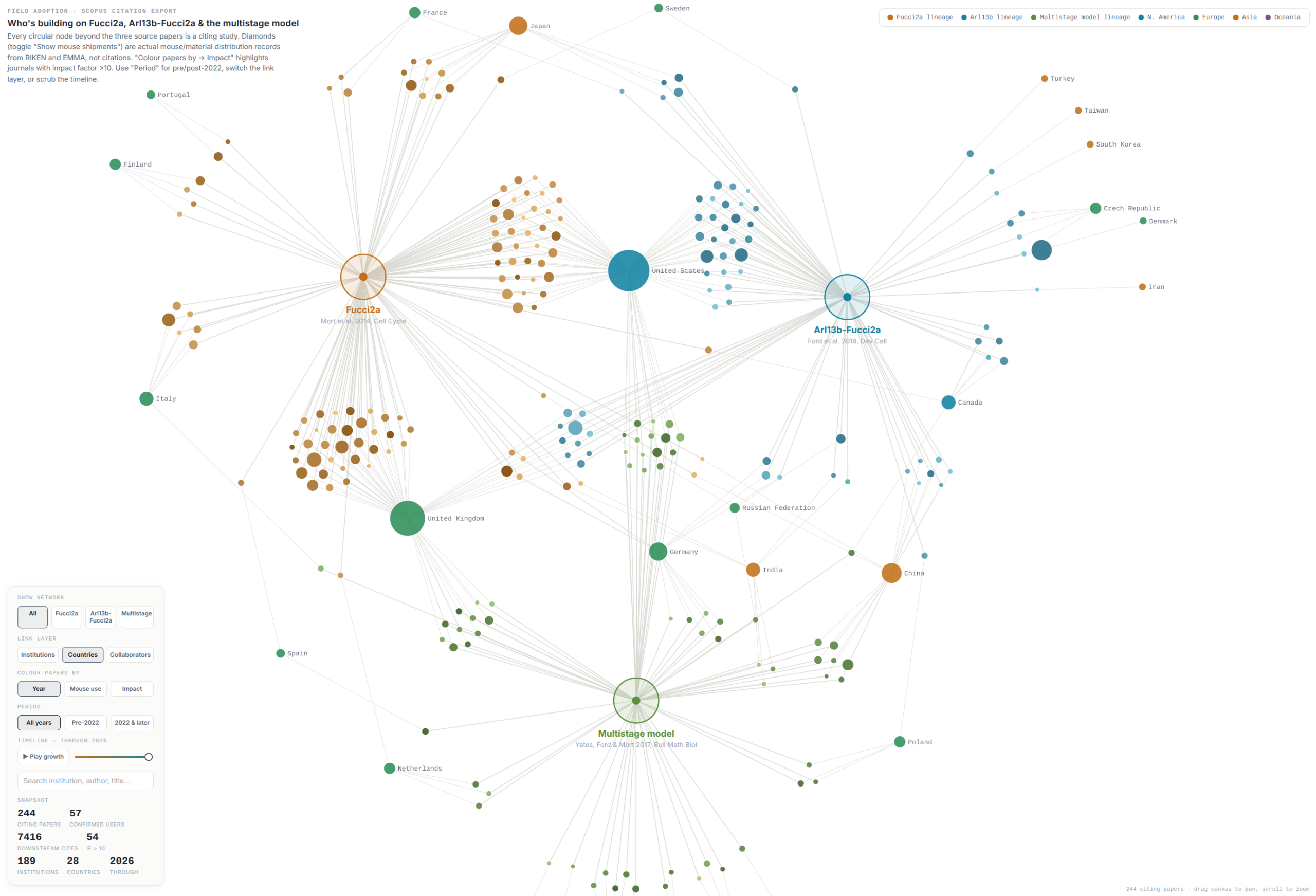
Interactive citation network coming soon — click image to enlarge
Key publications
Fucci2a: a bicistronic cell cycle reporter that allows Cre mediated tissue specific expression in mice. Mort RL, Ford MJ, Sakaue-Sawano A, Lindstrom NO, Casadio A, Douglas AT, Keighren MA, Hohenstein P, Miyawaki A, Jackson IJ. Cell Cycle. 2014;13(17):2681-96.
A Cell/Cilia Cycle Biosensor for Single-Cell Kinetics Reveals Persistence of Cilia after G1/S Transition Is a General Property in Cells and Mice. Ford MJ, Yeyati PL, Mali GR, Keighren MA, Waddell SH, Mjoseng HK, Douglas AT, Hall EA, Sakaue-Sawano A, Miyawaki A, Meehan RR, Boulter L, Jackson IJ, Mill P, Mort RL. Developmental Cell. 2018;47(4):509-523.
Genetically Encoded Tools for Optical Dissection of the Mammalian Cell Cycle. Sakaue-Sawano A, Yo M, Komatsu N, Hiratsuka T, Kogure T, Hoshida T, Ishidate Y, Toda S, Matsuda H, Miyawaki A. Molecular Cell. 2017;68(3):626-640.
An ILK/STAT3 pathway controls glioblastoma stem cell plasticity. Loftus AEP, Romano MS, Phuong AN, McKinnel BJ, Muir MT, Furqan M, Dawson JC, Avalle L, Douglas AT, Mort RL, Byron A, Carragher NO, Pollard SM, Brunton VG, Frame MC. Developmental Cell. 2024;59(24):3197-3212.
FuChi: A cell cycle biosensor expressing chicken for live imaging of cell cycle kinetics during avian development. Sudderick ZR, Briggs T, Van Kerckvoorde M et al. bioRxiv (in revision at PLoS Biology).
Collaborative publications
The following publications arose from active collaborations in which the Fucci2a platform was used alongside direct scientific contributions from the Mort Lab.
| An ILK/STAT3 pathway controls glioblastoma stem cell plasticity. Loftus AEP, Romano MS, Phuong AN, McKinnel BJ, Muir MT, Furqan M, Dawson JC, Avalle L, Douglas AT, Mort RL, Byron A, Carragher NO, Pollard SM, Brunton VG, Frame MC. Developmental Cell. 2024;59(24):3197-3212. |
| Epigenetic remodelling licences adult cholangiocytes for organoid formation and liver regeneration. Aloia L, McKie MA, Vernaz G, Cordero-Espinoza L, Aleksieva N, van den Ameele J, Antonica F, Font-Cunill B, Raven A, Cigliano RA, Belenguer G, Mort RL, Brand AH, Zernicka-Goetz M, Forbes SJ, Miska EA, Huch M. Nature Cell Biology. 2019;21(11):1321-1333. |
| Defining the Identity and Dynamics of Adult Gastric Isthmus Stem Cells. Han S, Fink J, Jörg DJ, Lee E, Yum MK, Chatzeli L, Merker SR, Josserand M, Trendafilova T, Andersson-Rolf A, Dabrowska C, Kim H, Naumann R, Lee JH, Sasaki N, Mort RL, Basak O, Clevers H, Stange DE, Philpott A, Kim JK, Simons BD, Koo BK. Cell Stem Cell. 2019;25(3):342-356. |
| Concerted cell divisions in embryonic visceral endoderm guide anterior visceral endoderm migration. Antonica F, Orietti LC, Mort RL, Zernicka-Goetz M. Developmental Biology. 2019;450(2):132-140. |
Selected independent studies using the Fucci2a platform
Papers from independent research groups (no co-authorship overlap with the Mort Lab) that have used the Fucci2a, Arl13b-Fucci2a, or related tools.
2026
Hypothyroidism impairs skeletal muscle regeneration after injury by altering myogenic and nonmyogenic pathways. Aguiari P et al. JCI Insight. 2026.
Reappraisal of profibrotic phenotype and cell-cycle state of renal tubular epithelium after ischemia–reperfusion injury. Fukaya D et al. PLOS ONE. 2026.
Precancerous niche remodelling dictates nascent tumour persistence. Skrupskelyte G et al. Nature. 2026.
Ets1-regulated endothelial-secreted factors promote compact myocardial growth and contribute to the pathogenesis of ventricular non-compaction. Wang L et al. Cardiovascular Research. 2026.
2025
Lifting regenerative barriers promotes epithelial cell fate plasticity supporting lineage conversion. Bejar MT et al. Nature Communications. 2025.
cSTAR analysis identifies endothelial cell cycle as a key regulator of flow-dependent artery remodeling. Deng H et al. Science Advances. 2025.
Injury-induced Clusterin+ cardiomyocytes suppress inflammation and promote regeneration in neonatal and adult hearts by reprogramming macrophages. Fan L et al. Cell Stem Cell. 2025.
Activity-dependent regulation of microglia numbers by pyramidal cells during development shape cortical functions. Kumaraguru S et al. Science Advances. 2025.
Zonal endothelial cell heterogeneity underlies murine renal vascular development. Luo PM et al. Angiogenesis. 2025.
Decoding cancer dormancy: integrative genomic, phenotypic and live-cell imaging analysis to reveal the hidden cancer cell reservoir. Porcelli G et al. Molecular Cancer. 2025.
Intronic RNAscope probes enable precise identification of cardiomyocyte nuclei and cell cycle activity. Yu Z et al. Communications Biology. 2025.
2024
Cell cycle–dependent centrosome clustering precedes proplatelet formation. Becker IC et al. Science Advances. 2024.
Protein translation rate determines neocortical neuron fate. Borisova E et al. Nature Communications. 2024.
Differential endothelial cell cycle status in postnatal retinal vessels revealed using a novel PIP-FUCCI reporter and zonation analysis. Liu Z et al. Angiogenesis. 2024.
Heterogeneity in oligodendrocyte precursor cell proliferation is dynamic and driven by passive bioelectrical properties. Pivoňková H et al. Cell Reports. 2024.
Exploring the principles of embryonic mammary gland branching morphogenesis. Satta JP et al. Development. 2024.
2023
Cell Cycle and Senescence Regulation by Podocyte Histone Deacetylase 1 and 2. Medina Rangel PX et al. Journal of the American Society of Nephrology. 2023.
Spatially coordinated cell cycle activity and motility govern bifurcation of mammary branches. Myllymäki SM et al. Journal of Cell Biology. 2023.
Profilin1 is required for prevention of mitotic catastrophe in murine and human glomerular diseases. Tian X et al. Journal of Clinical Investigation. 2023.
2022
DOT1L regulates chamber-specific transcriptional networks during cardiogenesis and mediates postnatal cell cycle withdrawal. Cattaneo P et al. Nature Communications. 2022.
Tubular cell polyploidy protects from lethal acute kidney injury but promotes consequent chronic kidney disease. De Chiara L et al. Nature Communications. 2022.
Lrig1 regulates the balance between proliferation and quiescence in glioblastoma stem cells. Ferguson KM et al. Frontiers in Cell and Developmental Biology. 2022.
Effect of disease progression on the podocyte cell cycle in Alport Syndrome. Frank CN et al. Kidney International. 2022.
SETD2 Haploinsufficiency Enhances Germinal Center–Associated AICDA Somatic Hypermutation to Drive B-cell Lymphomagenesis. Leung W et al. Cancer Discovery. 2022.
SyNPL: Synthetic Notch pluripotent cell lines to monitor and manipulate cell interactions in vitro and in vivo. Malaguti M et al. Development. 2022.
Differentiation of crescent-forming kidney progenitor cells into podocytes attenuates severe glomerulonephritis in mice. Melica ME et al. Science Translational Medicine. 2022.
FUCCI-Based Live Imaging Platform Reveals Cell Cycle Dynamics and Identifies Pro-proliferative Compounds in Human iPSC-Derived Cardiomyocytes. Murganti F et al. Frontiers in Cardiovascular Medicine. 2022.
Blocking cell cycle progression through CDK4/6 protects against chronic kidney disease. Osaki Y et al. JCI Insight. 2022.
Inhibition of DYRK1a Enhances Cardiomyocyte Cycling after Myocardial Infarction. Young A et al. Circulation Research. 2022.
2021
Loss of Endogenously Cycling Adult Cardiomyocytes Worsens Myocardial Function. Bradley LA et al. Circulation Research. 2021.
Tubular cell cycle response upon AKI: Revising old and new paradigms to identify novel targets for CKD prevention. De Chiara L et al. International Journal of Molecular Sciences. 2021.
Unexpected contribution of fibroblasts to muscle lineage as a mechanism for limb muscle patterning. Esteves de Lima J et al. Nature Communications. 2021.
Nutritional regulation of oligodendrocyte differentiation regulates perineuronal net remodeling in the median eminence. Kohnke S et al. Cell Reports. 2021.
LRIG1 is a gatekeeper to exit from quiescence in adult neural stem cells. Marqués-Torrejón MÁ et al. Nature Communications. 2021.
Vascular endothelial cell specification in health and disease. Marziano C et al. Angiogenesis. 2021.
A biomechanical switch regulates the transition towards homeostasis in oesophageal epithelium. McGinn J et al. Nature Cell Biology. 2021.
H3K27ac bookmarking promotes rapid post-mitotic activation of the pluripotent stem cell program without impacting 3D chromatin reorganization. Pelham-Webb B et al. Molecular Cell. 2021.
2020
Mutant EZH2 Induces a Pre-malignant Lymphoma Niche by Reprogramming the Immune Response. Béguelin W et al. Cancer Cell. 2020.
Abscission Couples Cell Division to Embryonic Stem Cell Fate. Chaigne A et al. Developmental Cell. 2020.
Altered G1 signaling order and commitment point in cells proliferating without CDK4/6 activity. Liu C et al. Nature Communications. 2020.
TBL1XR1 Mutations Drive Extranodal Lymphoma by Inducing a Pro-tumorigenic Memory Fate. Venturutti L et al. Cell. 2020.
2019
Single-Cell Analysis Reveals a Hair Follicle Dermal Niche Molecular Differentiation Trajectory that Begins Prior to Morphogenesis. Gupta K et al. Developmental Cell. 2019.
A stochastic framework of neurogenesis underlies the assembly of neocortical cytoarchitecture. Llorca A et al. eLife. 2019.
Oligodendrocyte Progenitor Cells Become Regionally Diverse and Heterogeneous with Age. Spitzer SO et al. Neuron. 2019.
2018
Hair follicle dermal condensation forms via FGF20 primed cell cycle exit, cell motility, and aggregation. Biggs LC et al. eLife. 2018.
Lgr5+ intestinal stem cells reside in an unlicensed G1 phase. Carroll TD et al. Journal of Cell Biology. 2018.
Endocycle-related tubular cell hypertrophy and progenitor proliferation recover renal function after acute kidney injury. Lazzeri E et al. Nature Communications. 2018.
Composite regulation of ERK activity dynamics underlying tumour-specific traits in the intestine. Muta Y et al. Nature Communications. 2018.
Genome-wide analyses reveal the IRE1a-XBP1 pathway promotes T helper cell differentiation by resolving secretory stress and accelerating proliferation. Pramanik J et al. Genome Medicine. 2018.
Fibroblast state switching orchestrates dermal maturation and wound healing. Rognoni E et al. Molecular Systems Biology. 2018.
Pyramidal cell regulation of interneuron survival sculpts cortical networks. Wong FK et al. Nature. 2018.
2016
Tissue-Specific Cell Cycle Indicator Reveals Unexpected Findings for Cardiac Myocyte Proliferation. Hirai M et al. Circulation Research. 2016.