Banner above. Left: Arl13b-Fucci2a labelled NIH3T3 cells (Matt Ford). Right H2B-Cerulean and EYFP labelled melanoblasts migrate in embryonic skin (Richard Mort).
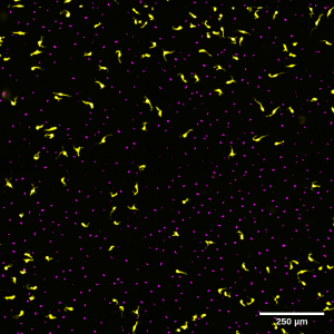
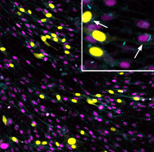
Live image of EYFP/H2BCerulean labelled melanoblasts migrating on the dorsal lateral pathway. (Richard Mort)
Live imaging of Qucci labelled NIH 3t3 cells (Tiernan Briggs)
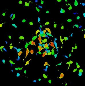
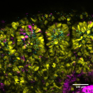
Depth coded stack of mouse melanoblasts as they begin to colonise a developing hair follicle at E14.5 (Richard Mort)
Live imaging of H2BCerulean-Fucci2a labelled NIH 3T3 nuclei. (Stephanie Wright)
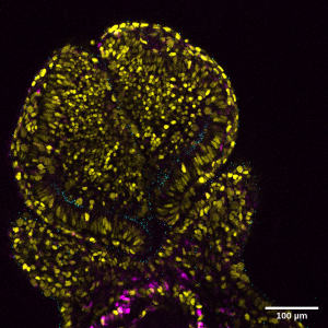
Live image of developing prosencephalon of an E8.5 mouse embryo labelled with Arl13bCerulean-Fucci2a. (Mill/Ford).
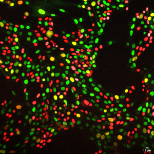
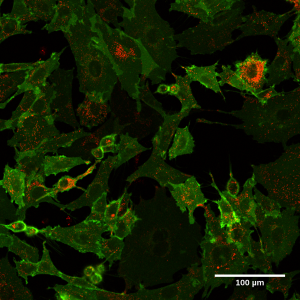
Live image of cKit-sfGFP/mCherry ‘timer tag’ labelled NIH 3T3 cells. (Olivia Harrison)
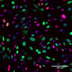
Live image of Arl13bCerulean-Fucci2a labelled NIH 3T3 cells. (Mill/Ford).
Melanocytes and macrophages in mouse tail skin (Emma Wilkinson)
Live image of developing somites of an E11.5 mouse embryo labelled with Arl13bCerulean-Fucci2a. (Mill/Ford).