Banner above. Left: Arl13b-Fucci2a labelled NIH3T3 cells (Matt Ford). Right H2B-Cerulean and EYFP labelled melanoblasts migrate in embryonic skin (Richard Mort).
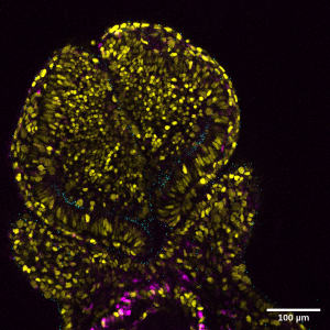
Live image of developing prosencephalon of an E8.5 mouse embryo labelled with Arl13bCerulean-Fucci2a. (Mill/Ford).
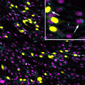
Melanocytes and macrophages in mouse tail skin (Emma Wilkinson)
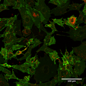
Live image of cKit-sfGFP/mCherry ‘timer tag’ labelled NIH 3T3 cells. (Olivia Harrison)
Live imaging of Qucci labelled NIH 3t3 cells (Tiernan Briggs)
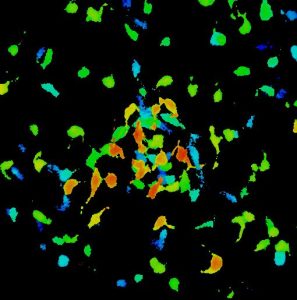
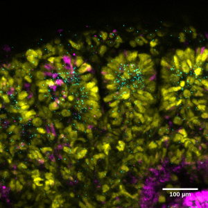
Depth coded stack of mouse melanoblasts as they begin to colonise a developing hair follicle at E14.5 (Richard Mort)
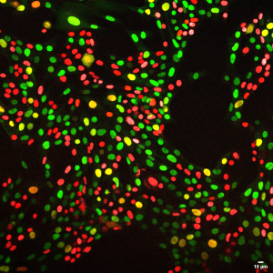
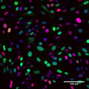
Live imaging of H2BCerulean-Fucci2a labelled NIH 3T3 nuclei. (Stephanie Wright)
Live image of developing somites of an E11.5 mouse embryo labelled with Arl13bCerulean-Fucci2a. (Mill/Ford).
Live image of Arl13bCerulean-Fucci2a labelled NIH 3T3 cells. (Mill/Ford).
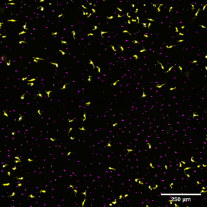
Live image of EYFP/H2BCerulean labelled melanoblasts migrating on the dorsal lateral pathway. (Richard Mort)